
Researchers from the Massachusetts Institute of Technology (MIT) developed a noninvasive patch that can generate excellent, real-time images of organs for a period of two days.
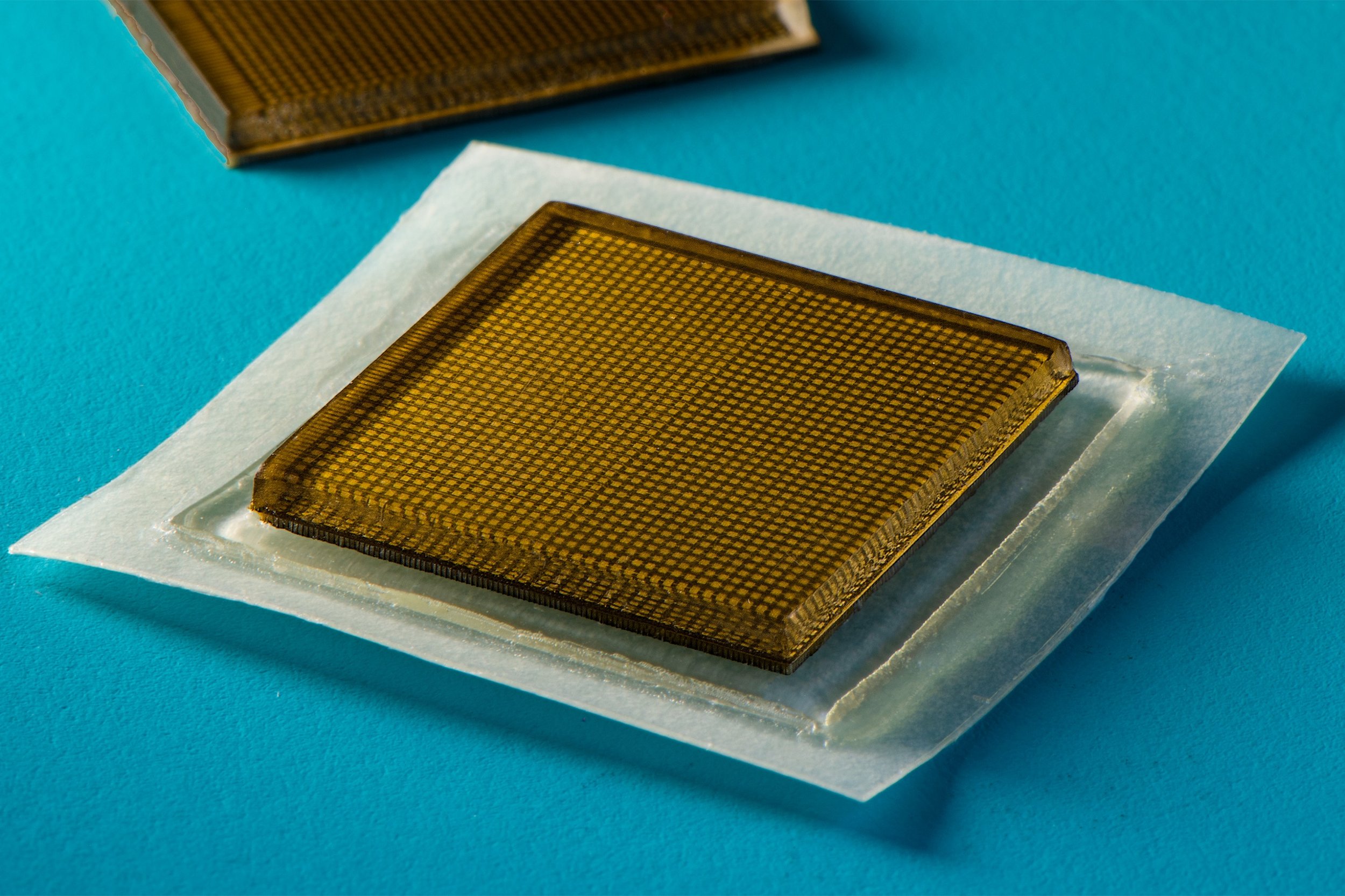
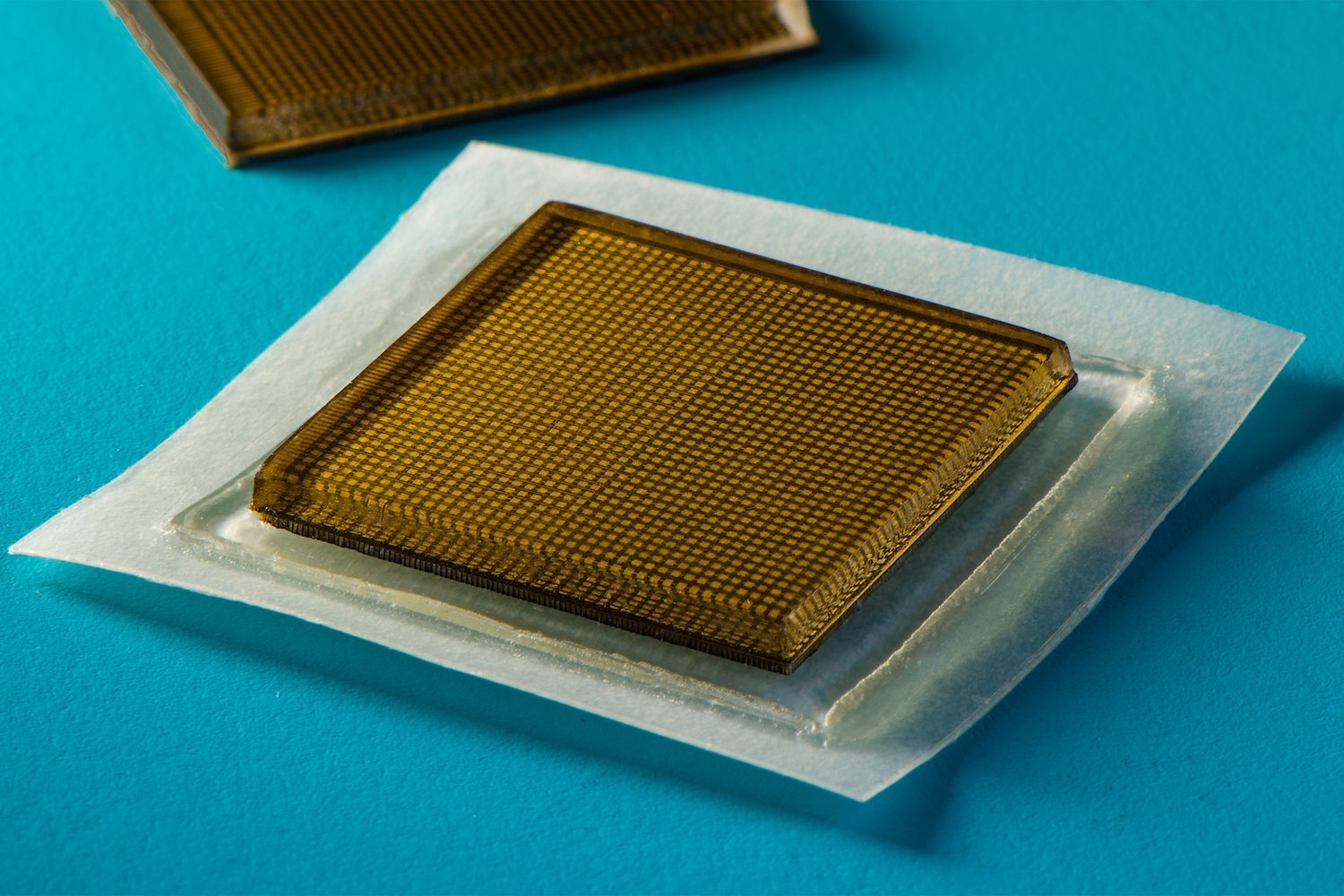
The MIT developed patch capable of producing real time images of organs within the body - Image Credit Felice Frankel via EurekAlert
Bringing ultrasound technology to your local drug store
Up until now, medical specialists have been using conventional ultrasound imaging to get a look at our organs without the need for keyhole surgery or other more invasive procedures.
Contemporary ultrasound imaging comes with a significant disadvantage, namely that it requires large, expensive equipment. This type of equipment is usually only reasonable in the context of a doctor's office or hospital.
However, a team of MIT researchers aims to bring this technology much closer to people with a new high-tech patch that could be widely available in the future, perhaps even in your local drug store.
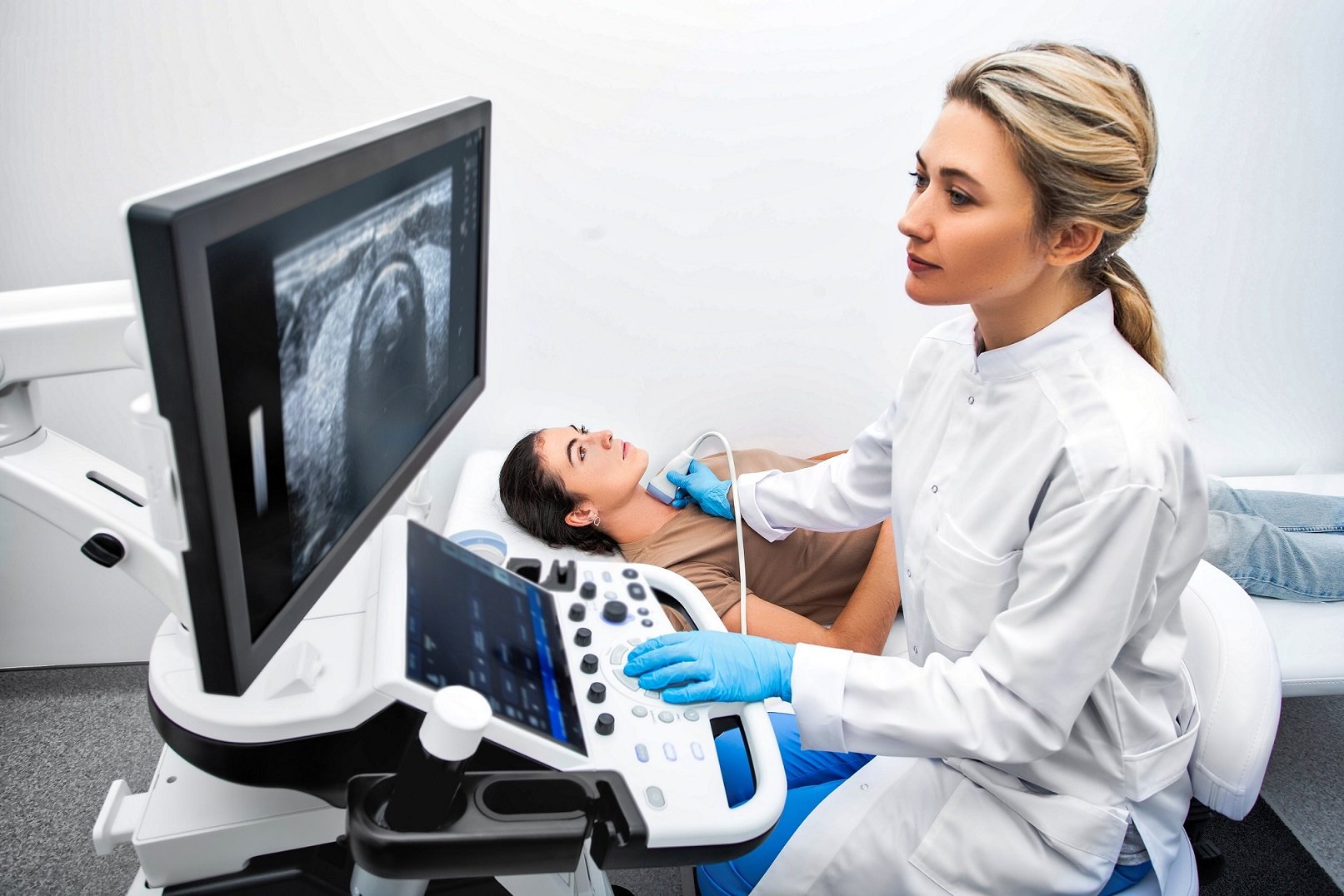
Conventional ultrasound technology - (Image Credit: Peakstock via Shutterstock / HDR tune by Universal-Sci)
A patch that can see inside your body
The team developed a device, the size of a stamp, that is capable of providing uninterrupted ultrasound imaging of organs for two days.
Applying the stamp-sized gadget to research participants yielded real-time, high-res pictures of deeper organs, such as the stomach, heart, and lungs.
Whilst wearing the patch, participants engaged in a variety of activities, such as running, standing, sitting, and bicycling; the patches remained firmly attached while recording changes in the underlying organs.
From the imagery generated by the patches, the team could observe the changing width of major blood vessels when participants were standing versus being seated.
The patches were also capable of capturing information about deeper organs, such as how the stomach distends and then shirks back as volunteers drink fluids, as well as the heart transforming its shape as it exerts during exercise.
Some participants lifted weights for their part of the testing. While lifting weights, the researchers were able to discern bright patterns in underlying muscles, indicating brief microdamage.
The patch is capable of generating high-resolution images over a longer duration by pairing an elastic adhesive layer with a rigid array of transducers.
According to MIT graduate student Conghe Wang, this setup allows the device to conform to the skin while preserving the relative location of transducers to develop more detailed and clearer images.
Making it wireless
In its current iteration, the patches still require a wired connection to a device that can convert the reflected sound waves into viewable images.
Although the patches have benefits even in their current form compared to conventional ultrasound equipment, the team is working on making them operate wirelessly.
Making the patches wireless would mean that patients could take them home from the hospital or doctor's office or possibly even purchase them at a drug store.
MIT professor, Xuanhe Zhao, stated that the team is foreseeing a set of patches that can be put on different locations of your body while communicating with your smartphone.
In principle, it would be possible for software on your phone to analyze the images on demand, making the patches a very potent medical tool.
Xuanhe Zhao: "We believe we've opened a new era of wearable imaging: With a few patches on your body, you could see your internal organs."
The team presented their findings in a paper published in the peer-reviewed science journal Science, listed below for those interested in more details.
Sources and further reading:
If you enjoy our selection of content consider subscribing to our newsletter (Universal-Sci Weekly)
FEATURED ARTICLES: