Antibiotics are one of the wonders of modern health science, saving millions of lives every year. But, unfortunately, they are starting to fail. We have been sitting on a ticking timebomb since the rise of antibiotic resistance.
One of the reasons that some bacteria are harder to terminate with antibiotics is the protective layer that envelops them.
Now, an international collaboration of scientists managed to create the most detailed images ever of a living E.ocli bacterium, revealing some very useful information that may help us in the fight against antibacterial resistance.

It is estimated that antibiotic resistance may claim as many as ten million lives - per year - by 2050. - Image Credit: Saiful52 via Shutterstock / HDR tune by Universal-Sci
The defensive prowess of gram-negative bacteria
Bacteria equipped with a benefitting protective outer membrane shielding them against antibiotics are known as 'gram-negative bacteria.'
According to Bart Hoogenboom, corresponding author of the study, this outer membrane provides a tremendous antibiotic barrier. It is one of the main contributors that make infectious bacteria resistant. He goes on to explain that scientists are uncertain how exactly this barrier is assembled—making it an excellent target for their study.
The study
To get a better understanding of the aforementioned membrane, the scientists started working with a well-known bacterium called Escherichia coli (or e.coli).
They took a truly minuscule needle (with a diameter of only a few nanometers) and ran over the living bacterium to 'feel' its shape. Because the tip of this needle was so small, the researchers were able to 'feel' detect minuscule structures on the bacterium's surface.
This method generated the most detailed images of a living bacterium ever, uncovering some surprising information.
The results
The image revealed that gram-negative bacteria's shielding 'envelope,' as it is sometimes called, is rather complex. It consists of dense meshes of protein building blocks alternated by patches that appear to be protein-free.
It turns out that these 'protein-free spots are filled with so-called glycolipids. In cells, their role is to support the stability of the cell membrane. They are vital in the connections that allow cells to attach to one another to form tissues. In this case, the glycolipids keep the protective layer firm.
Furthermore, the scientists discovered that a distinct type of pimple-like patch appeared when sections of the membrane were inverted on this particular bacterium as a result of mutations.
In this instance, the existence of these mutations correlated with a heightened susceptibility to an antibiotic known as 'bacitracin. Normally this specific antibiotic is only efficacious in fighting gram-positive bacteria.
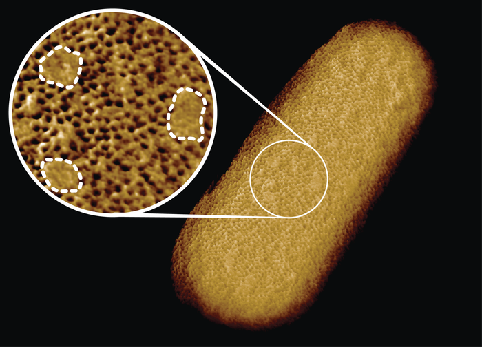
The image exposed the patchy surface of the outer membrane. The smooth, protein-free patches are lablled by dashed lines in this image - Image Credit: Benn et al. UCL
Potential weak spots
Georgina Benn, responsible for the microscopy for this study, explained that their images paint a different picture than currently found in textbooks. Instead of proteins distributed in a distorted manner, the team found lipid patches separated from protein-rich networks akin to oil separating from water.
With this new way of looking at the outer membrane, scientists can now start investigating what these structures mean for membrane integrity, function, and, perhaps most importantly, antibiotic resistance.
According to Hoogenboom: we can now see how membrane proteins establish a network that stretches around the whole exterior of the bacteria, leaving tiny gaps for spots that lack protein.
This indicates that the 'armor' of these bacteria may not be impenetrable after all. As it isn't evenly distributed over the entire surface. It may have weaker spots that can be targeted by specialized antibiotics.
Hopefully, this study will eventually lead to scientists being able to use the weak spots in the membrane to combat gram-negative bacteria.
The scientists published their findings in the scientific journal: Proceedings of the National Academy of Sciences. It is listed below for those interested.
Sources and further reading:
Will AI rescue us from a world without working antibiotics? (Universal-Sci)
Phase separation in the outer membrane of Escherichia coli (Proceedings of the National Academy of Sciences)
Gram-negative bacteria (CDC)
E. coli (Mayo Clinic)
The next 'pandemic' is antibiotic resistance, a ticking time bomb, and we are doing nothing to stop it (Universal-Sci)
Glycolipid (Biology Online)
If you enjoy our selection of content, consider subscribing to our newsletter - (Universal-Sci Weekly)
FEATURED ARTICLES: